Ultrasound Structured Reporting Software
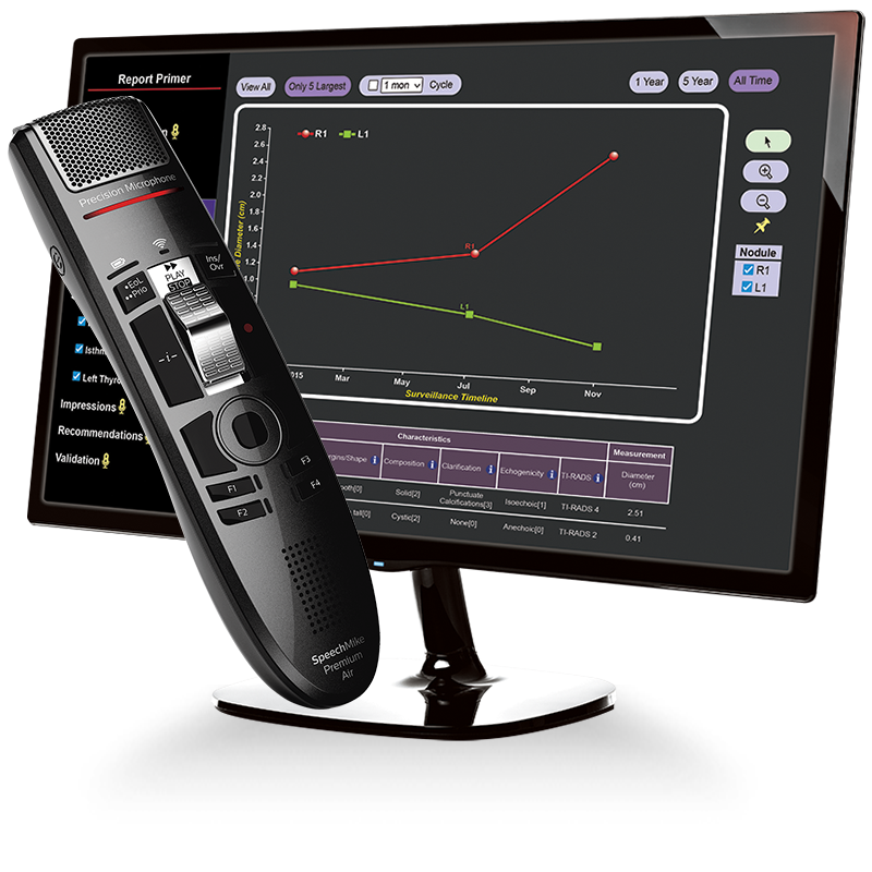
9i Voice Activated, Hands-Free Digital Structured Reporting
…Old English Proverb
“Fast is Good, Faster is Better, Fastest is Best”…Old Radiology Proverb
CLICKVIEW 9i: Hands-Free Ultrasound Structured Reporting Software
All contemporary computer-based reporting systems, such as structured templates and speech recognition systems, rely on the computer mouse and keyboard to navigate and scroll the display screen to enter text and data. This creates significant distractions from the radiologist’s focus on describing and diagnosing the image findings on the screen. The mouse and keyboard slow report generation and are an annoying interruption of the radiologist’s workflow.
CLICKVIEW 9i’s innovative Voice Activation Ultrasound Structured Reporting Software user interface uses speech recognition to eliminate the need for the mouse and keyboard as text and data input devices for most reports. The user dictates the report in natural language, and the Smart Engine automatically places the text in the appropriate anatomical section of the report. Context-sensitive Macros are inserted into the report using simple voice commands. User experience is enhanced by increased efficiency, faster report generation, and a more intuitive workflow.
CLICKVIEW Corporation pioneered the development and application of Structured Reporting in the late 1970s and early 1980s. At the time, there was an urgent need to report new data-dense Gray Scale Obstetrical (OB) Ultrasound reports more clearly than standard text-only reports. Simultaneously, there was an urgent need to reduce the high medical transcription costs associated with generating the reports. The solution was to use emerging personal computer technology as a platform to create multimedia graphical reports.
To better understand the development of ultrasound reporting software in radiology, it is helpful to briefly review how structured reporting was developed in radiology, specifically for OB ultrasound.
Figures 1 and 2 are the same OB Ultrasound report. Figure 1 is a conventional dictated all-text report, and Figure 2 is a multimedia graphical report utilizing a trend chart (similar to Professor Campbell’s original publications) to display a fetal growth curve and tables graphically organizing relevant biometric data and calculations for Estimated Gestational Age (GA) and Fetal Weight (FW), along with other findings.
Which is easier and faster to understand the exam findings?
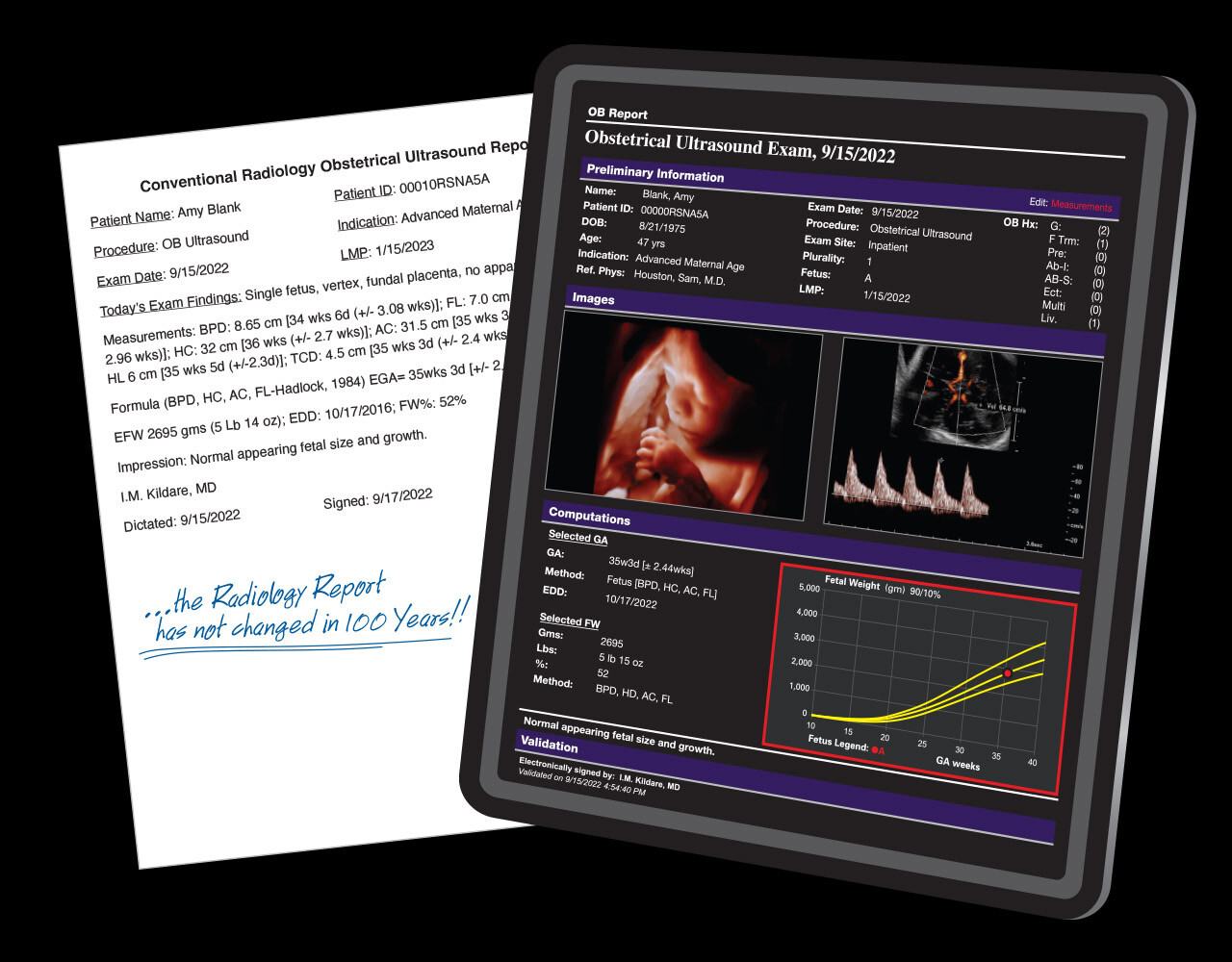
Bistable OB Ultrasound Data: Biparietal Diameter (BPD) and Abdominal Circumference (AC).
Stuart Campbell, DSc FRCPEd FRCOG FACOG, considered the father of obstetrical ultrasound, published the earliest papers on fetal biometry using bistable scan devices, which displayed images in black and white video without gray shading. Professor Campbell published articles demonstrating BPD for the first time (1968), BPD growth chart (1969), IUGR BPD fetal growth chart (1971), and the first AC chart in 1975. Dr. Campbell’s obstetrical ultrasound reports consisted of one or both measurements as the basis for Gestational Age (GA) and Abdominal Circumference (AC) growth charts. Early OB reports, using only BPD.
Gray Scale and Real Time Ultrasound
Gray scale imaging ultrasound scanners were first widely commercially available in the US in the mid-70s and produced high-quality grayscale images, which, for the first time, accurately visualized fetal soft tissues and enabled the development of additional fetal biometry. The improved accuracy of grayscale images of fetal biometry was the basis for the use of statistical analysis of the primary fetal biometric measurement data for the determination of Gestational Age (GA), Fetal Weight (FW), Estimated Delivery Date (EDD), and other. Real-time ultrasound scanners were added to the profusion of data and calculations to quantify normal and abnormal fetal growth with new anatomical, cardiac, vascular, and Doppler parameters.
The PC
In parallel to the evolution of OB ultrasound, the personal computer industry was in its infancy. With the release of MS-DOS in 1980, the IBM PC in 1981, and PC-specific programming languages, program development became accessible and affordable to anyone with an interest or a need. Earlier pre-DOS versions of the original CLICKVIEW program were ported to the MSDOS platform, and the program’s development was accelerated. Within the next two years, the earliest CLICKVIEW ultrasound structured reports incorporating fetal growth charts and tables were sent to referral physicians on a routine basis. Report generation time was reduced to minutes, and transcription costs were reduced by about 90%.
Need to See the Data
The surge of fetal biometric data expanded the need to communicate dense OB data reports more efficiently than conventional text reports. Extensive multi-media displays of fetal data were developed, including charts, tables, illustrations, images, cine clips, and a wide array of reporting macros to describe the broad spectrum of findings displayed by the greyscale and real-time technology.
In a short time, however, the burden of graphic display exceeded the capability of contemporary PCs to process reports efficiently. This stifled the interest of general radiologists, other than those in Ultrasound, in adopting structured reporting more widely. In addition, the profound cultural inertia of dictation as the conventional way of report generation contributed to the resistance to using a mouse and keyboard instead of dictation in a speech recognition system.
Nonetheless, data display concepts developed for obstetrical reports and the power of advanced multimedia programming languages quickly migrated to general ultrasound reports of the abdomen, Doppler studies of the vascular system, thyroid nodules, tumor tracking, and other reports.
There is a reason we are referred to as
“The radiology structured reporting company”
35+
Years Structured Reporting Experience
25
Million Reports
20+
Years Web Architecture
~2.5
~1,500
Active Weekly Users