“The history of sonography in Obstetrics and Gynaecology dates from the classic 1958 Lancet paper of Ian Donald and his team from Glasgow. Fifty years on it is impossible to conceive of practicing Obstetrics and Gynaecology without one of the many forms of ultrasound available today” (1)
In his excellent, “A Short History of Sonography in Obstetrics and Gynaecology,” (1) Professor Stuart Campbell provides us with a remarkable and unique, first-person panoramic view of the evolution of OB/GYN ultrasound technology, to which he was also a seminal contributor, over the last half of the 20th century. The following summary offers key highlights of the article as an overview. References and literature citations are listed in the original article.
Early History
To begin with, Campbell reminds us that the first publication by Ian Donald in 1958 of bistable images of the fetus and fetal head rests on foundational basic scientific contributions dating back 150 years to the description of ‘phase shifting’ by Thomas Young in 1801, as well as Christian Doppler’s 1842 description of the Doppler effect in describing the motions of the stars, and the later work of Pierre Curie (5) describing the piezoelectric effect in 1880, and others.
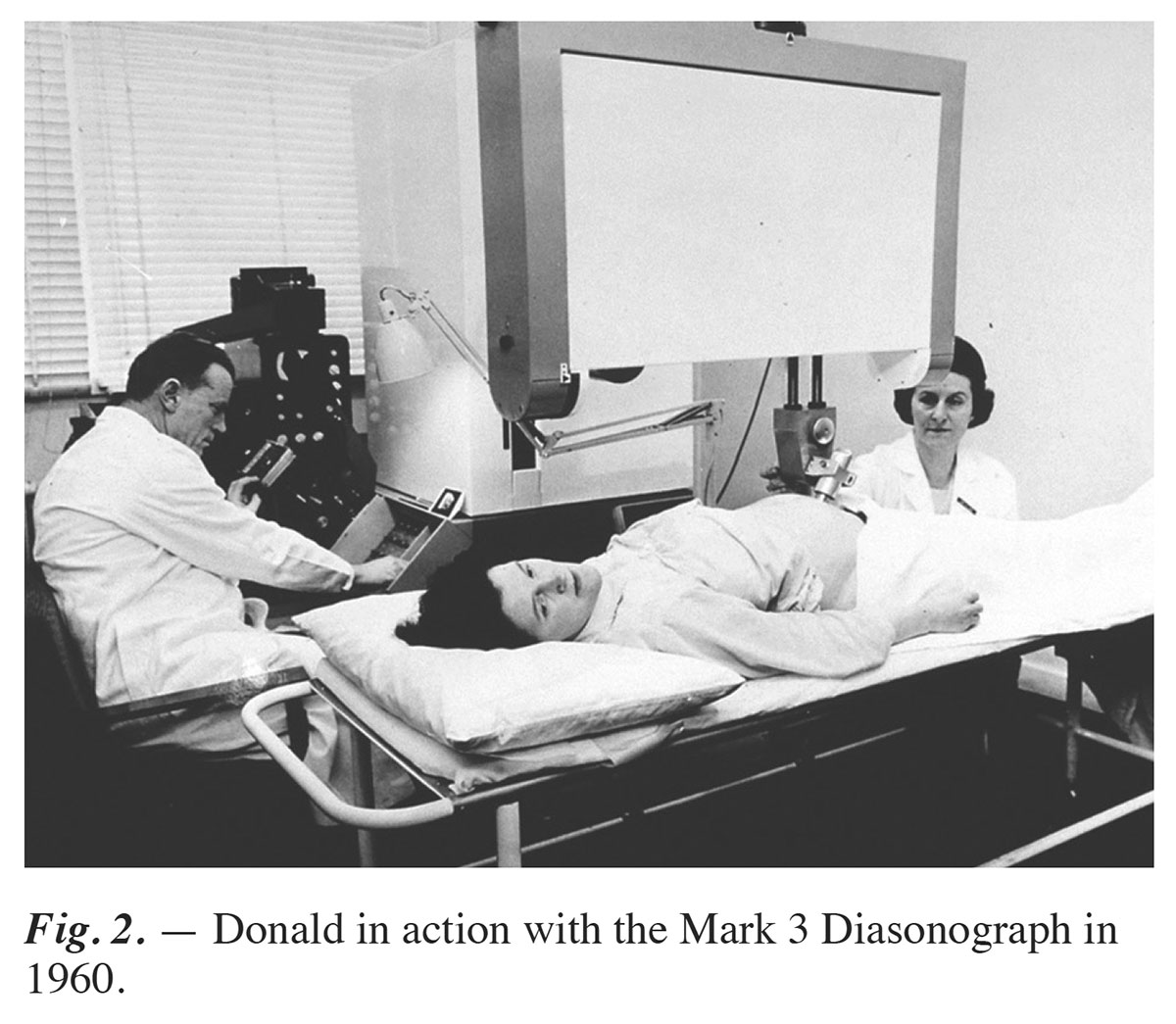
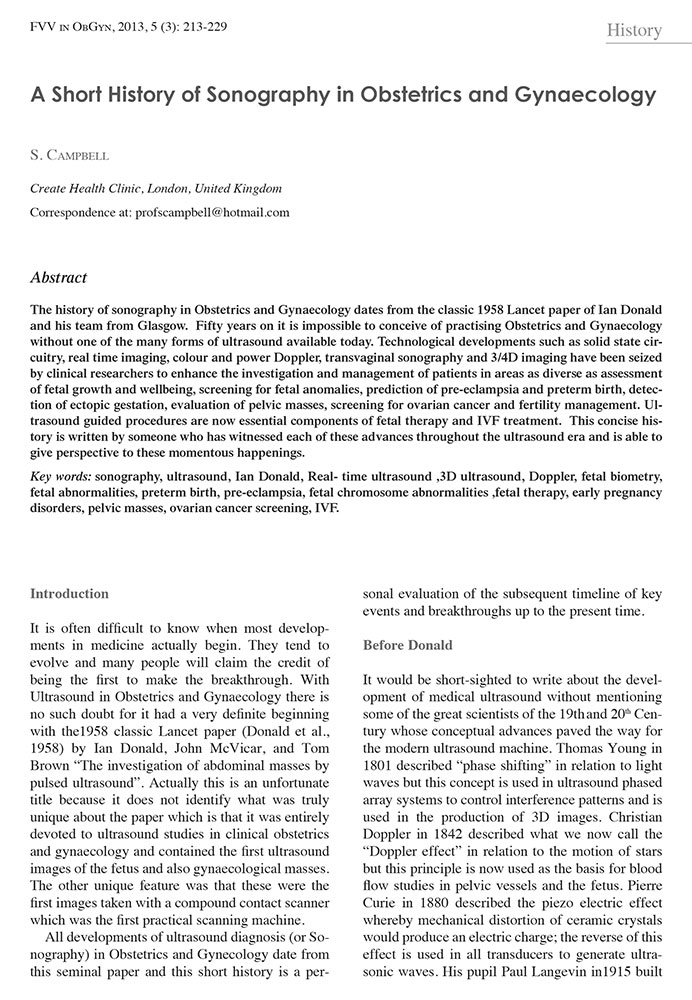
The basic science led to the development of the first ultrasound ‘water bath’ technology for applications to human anatomy by Douglas Howry in 1952, including gall stones, the adult heart and breast tumors, and other human anatomy. Six years later, Donald developed the first compound 2D contact scanner and published the first ultrasound image of a fetal BPD.
Bistable clinical applications in OB/GYN ultrasound moved over the next twenty years from descriptive OB/GYN anatomy such as placental location and congenital abnormalities to assessing fetal growth.
Significant Advances
Campbell’s seminal 1969, 1971, and 1975 bistable fetal biometry growth charts of BPD and AC of normal and abnormal fetal growth were foundational for assessing gestational age, fetal weight, IUGR and predicting EDD. Huch Robinson’s description in 1973 of the fetal crown-rump length dating of the embryo anchored early sonographic dating methods from 7-16 weeks within +/- 5 days.
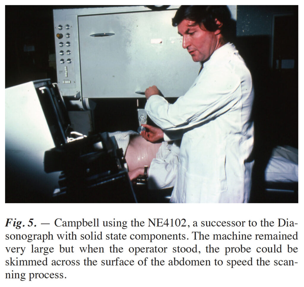
Scan converter technology, which converted the display of the compound images in scales of grey for different tissues, became widely available in commercial scanners in the mid-1970s and immediately allowed users to see more detailed anatomy and develop extensive biometry charts for the fetus.
The Floodgates Are Open
Ten years later, the introduction of high-resolution real-time scanning technology by Acuson and the development of color-flow Doppler opened the floodgates to an enormous proliferation of research describing the anatomy and pathology of the fetus and pregnancy as well as attempts at intrauterine interventions for congenital defects.
Today, OB/GYN ultrasound is the standard of care worldwide to monitor low-risk pregnancies and to detect and manage high-risk pregnancies.
- Campbell, S. “A Short History of Sonography in Obstetrics and Gynaecology,” FVV in ObGyn, 2013, 5 (3): 213-229